Key Pointers
- A heart perfusion test shows how well blood flows to your heart muscle at rest and during stress.
- “Normal” results mean the heart muscle is getting adequate blood supply.
- “Abnormal” results may indicate restricted blood flow (ischemia) or prior heart muscle injury.
- These results help your cardiologist plan next steps, such as medication, angiogram, or stenting.
What Is a Heart Perfusion Test?
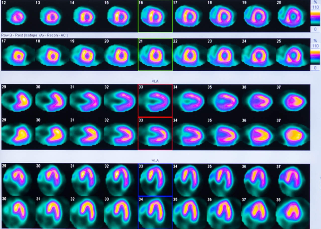
A Heart Perfusion Test, often called a Myocardial Perfusion Imaging (MPI) scan, helps doctors assess blood flow to the heart muscle. It’s usually done after a stress test, where the heart is “stressed” through exercise or medication, followed by imaging using a small amount of radioactive tracer.
The tracer travels through the bloodstream and highlights how much blood reaches different parts of the heart. A special camera then creates images showing the areas that are well-supplied versus those receiving less blood.
Why Doctors Recommend This Test
Your cardiologist may suggest a heart perfusion test if you:
- Experience chest pain, breathlessness, or unexplained fatigue
- Have abnormal results from an ECG, echocardiogram, or treadmill stress test
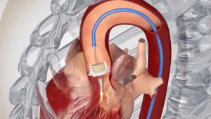
- Previously had angioplasty or bypass surgery and need follow-up evaluation
- Are being assessed before major surgery
This test provides more detail than a regular stress test because it visualises how each segment of the heart muscle is perfused, helping doctors determine whether blockages in the coronary arteries are affecting heart function.
What Are the “Normal” Results
When the report says your heart perfusion test is normal, it means:
- Blood flow to all parts of the heart muscle is adequate during both rest and stress.
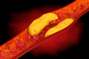
- There are no significant blockages in the coronary arteries affecting blood supply.
- The heart is pumping efficiently, with normal wall motion and uniform uptake of the tracer.
A normal result reassures both patient and doctor that the heart is receiving enough oxygen-rich blood even during exertion.
What “Abnormal” Results May Indicate
An abnormal perfusion result doesn’t automatically mean you’ve had a heart attack, but it signals an area of concern that needs further assessment.
There are two main patterns:
1. Reversible Defect (Ischemia)
- Blood flow is reduced during stress but normal at rest.
- Suggests that one or more coronary arteries are partially narrowed.
- Indicates that the heart muscle is still alive but under stress when blood demand increases.
2. Fixed Defect (Infarction or Scar)
- Blood flow is reduced both at rest and during stress.
- Suggests that the heart muscle in that area has been permanently damaged, often from a previous heart attack.
Sometimes the scan may show mixed defects, where part of the heart muscle has scarring and another part shows stress-induced ischemia.
Summary of Perfusion Test Results
|
Result Type
|
Blood Flow at Rest
|
Blood Flow During Stress
|
What It Means
|
|---|---|---|---|
|
Normal
|
Normal
|
Normal
|
Adequate blood supply to the heart; no significant blockage
|
|
Reversible Defect (Ischemia)
|
Normal
|
Reduced
|
Narrowed coronary arteries; possible restricted blood flow during exertion
|
|
Fixed Defect (Scar)
|
Reduced
|
Reduced
|
Previous heart muscle damage, such as from an old heart attack
|
|
Mixed Defect
|
Partial
|
Reduced
|
Combination of scarring and reversible narrowing
|
This simplified summary helps patients understand how cardiologists interpret MPI results in relation to heart blood flow and muscle health.
What Affects the Accuracy of Results
Several factors can influence test findings, including:
- Body build or breast tissue that may interfere with image quality
- Medications that affect heart rate or blood pressure
- Incomplete stress response, if exercise or medication didn’t raise heart rate sufficiently
- Technical artefacts due to movement during imaging
For this reason, results should always be reviewed alongside your symptoms, ECG, echocardiogram, and medical history.
What Happens After an Abnormal Result
If the scan shows reduced blood flow (ischemia), your cardiologist will discuss next steps, which may include:
- Coronary angiogram – to directly visualise the narrowed artery
- Angioplasty and stenting – if a blockage is found and needs to be opened
- Optimising medication – such as anti-anginal drugs or cholesterol-lowering therapy
- Lifestyle modification – including smoking cessation, diet, and exercise guidance
The goal is not only to treat the narrowed artery but also to protect the rest of your heart from future events.
Frequently Asked Questions
1. What is the difference between a stress test and a perfusion scan?
A treadmill stress test records ECG changes, while a perfusion scan adds imaging to show actual blood flow in the heart muscle.
2. How long does the test take?
It typically takes a few hours, as imaging is done twice, once at rest and again after stress, to compare perfusion.
3. Can an abnormal scan improve later?
Yes. With appropriate treatment such as medication or stenting, blood flow can be restored, and repeat imaging may show improvement.
4. Is the test safe?
The radioactive tracer dose is small and considered safe for most patients. Your cardiologist will evaluate suitability based on your health and medical history.Also, You should stay away from pregnant lady and infant for the next 24 hours.
Take the Next Step, Book an Appointment
If you’ve recently had a heart perfusion test and are unsure about what the results mean, it’s best to discuss them with a cardiologist experienced in coronary imaging and interventional cardiology.
At Heart Specialist International, our team provides comprehensive cardiac evaluation, from non-invasive imaging such as myocardial perfusion scans to interventional procedures like angioplasty and stenting when needed.
- Call +65 6962 1287
-
Mount Elizabeth Novena Specialist Centre,
#07-41, 38 Irrawaddy Road, Singapore - Book an Appointment